
 Menu
Menu

Menu
Explore the Brain from Different Angles

MATERIAL MADE JUST FOR YOU:
HOW DO INTERACTIVE IMAGES WORK?
Watch the video and see how simple and quick it is to understand how all our interactive tools work. Notice that when you hover the mouse over a specific region, it highlights, and an identification box appears with the name of the indicated area.
In addition, you can zoom in on regions of interest, allowing you to explore even very small areas and providing a more interactive and engaging experience while studying Neuroanatomy. Enjoy!
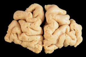
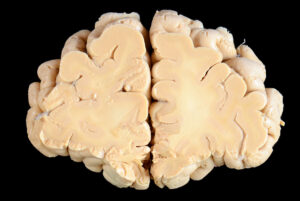
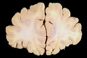
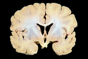
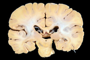
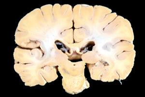
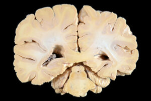
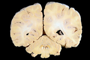
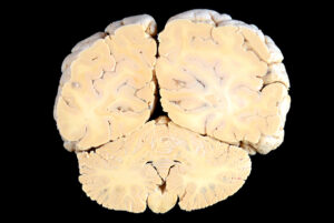
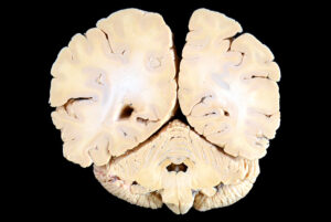
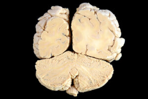
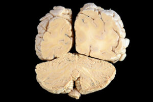
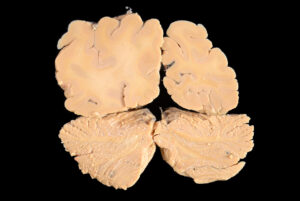
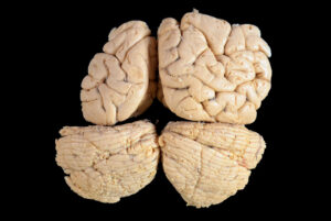
SECTIONAL ANATOMY OF THE BRAIN - CORONAL SECTIONS
High-resolution photos with identified structures

Coronal Sections made with 5mm thickness.
Also check out this material in its interactive form and get more information about the structures and regions of the encephalon!!!











SECTIONAL ANATOMY OF THE ENCEPHALON - AXIAL SECTIONS
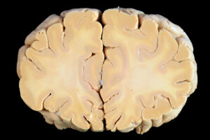
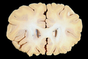
Study the Brain through Axial (Transverse) Sections
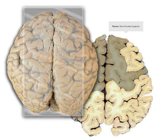
Axial Sections made with 5mm thickness.
Study through properly captioned and revised photos like a conventional atlas and/or interact with our interactive content in a more dynamic way.
MATERIAL DEVELOPED BY STUDENTS FOR STUDENTS!
This project was developed at Neuromorphology Laboratory of the Department of Structural and Functional Biology of the Institute of Biosciences of Botucatu - UNESP, and aims to facilitate the learning of Neuroanatomy.

Our Team

Science Teacher/Web Designer

Project Coordinator Professor;
Currículo Lattes

Collaborating Professor/Reviewer;
Currículo Lattes

Collaborating Professor/Reviewer;
Currículo Lattes
Photography Area
Sílvio Geraldo dos Santos Júnior –
Photography Area
Copyright © 2026 Ei_Atlas | Powered by Ei_Atlas